g.HIamp is a high-performance biosignal amplifier with 256 channels for invasive and non-invasive measurements of the brain that is FDA cleared and CE approved. g.HIamp provides excellent signal resolution and a wide input sensitivity to record EEG, ECoG, ECG, EMG, and EOG without any saturation. External body sensors can also be connected. Internally, signal processing is performed with the fastest floating-point DSP and a sophisticated real-time Kernel. The amplifier relies on a very high oversampling to reduce the noise as much as possible by averaging samples.
256 channels can be analyzed in real-time with g.HIsys Highspeed Processing for Simulink software. This provides faster and more accurate control of BCI systems that use Common Spatial Patterns (CSP). g.HIamp is equipped with 16 digital trigger channels and a HOLD input for artifact suppression (e.g. during electrical or magnetic stimulation).
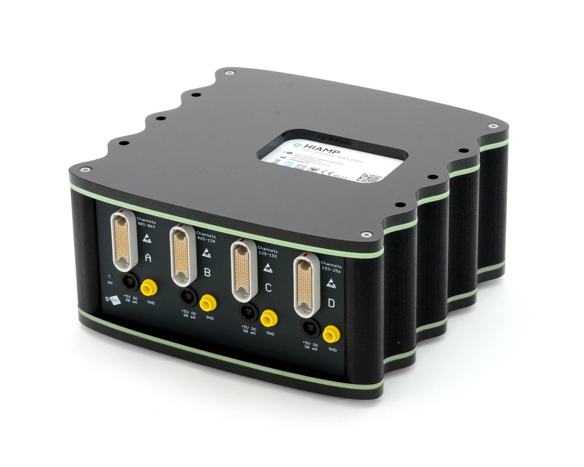
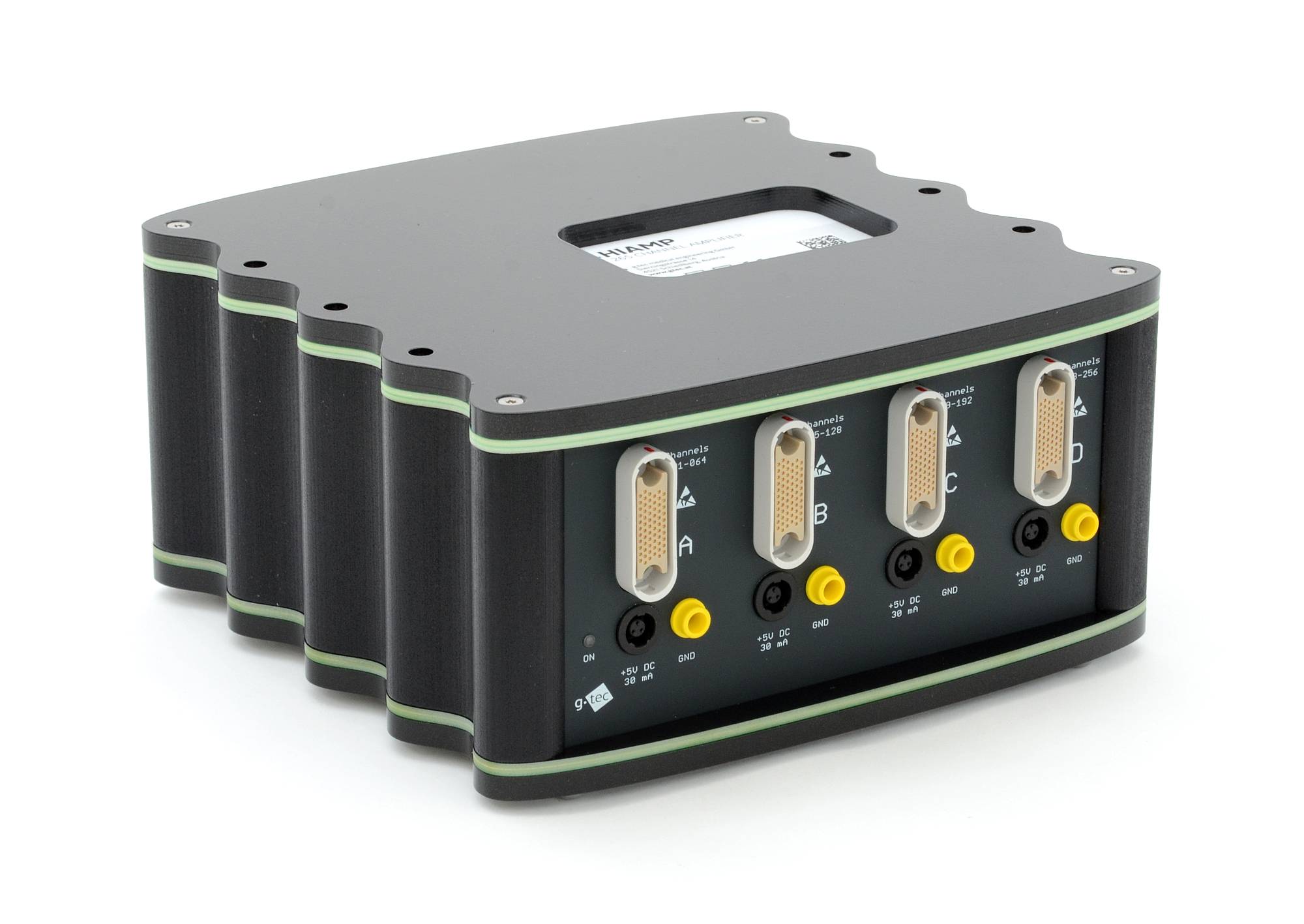
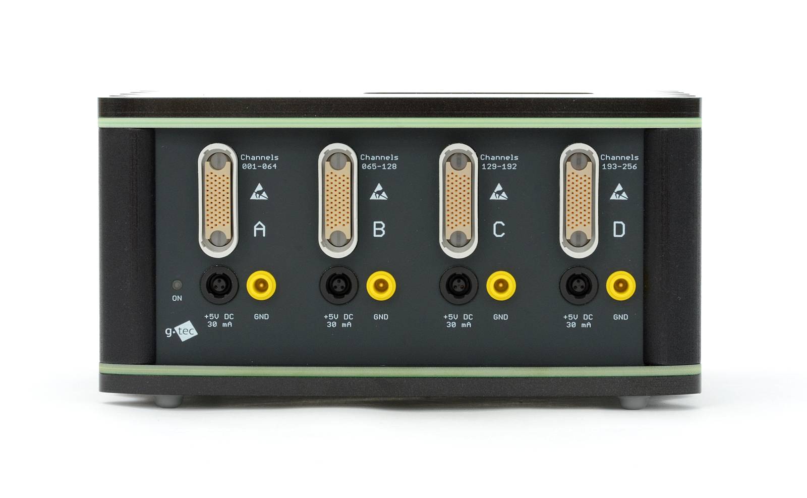
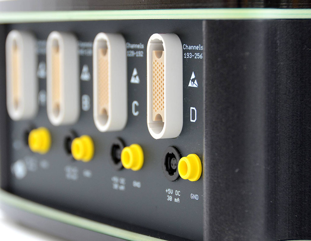
g.HIamp EEG amplifier has 80, 144 or 256 channels and can be used with passive or active EEG, ECoG, ECG, EMG and EOG electrodes. The difference between these options is just the electrode connector box. Each block of 64 channels is connected via a multi-pole medical safety connector to the electrode box. For ECoG grids and strips, special interface connectors are available too.
| High-performance EEG/EMG/ECG/... amplifier | |
| Fully integrated into g.tec Suite 2020 software environment for real-time or offline analysis | |
| 80/144/256 DC coupled wide-range input channels, able to record any type of signal (EEG, ECoG, EMG, EOG/spikes, connected various sensors) | |
| 256 channels perfectly synchronized with 24 Bit resolution | |
| High-gamma up to 1 kHz | |
| Integrated impedance measurement for active and passive electrodes | |
| Simultaneous TMS/tDCS/fNIRS recordings possible | |
| Easy configuration and setup with g.HIsys Professional for Simulink | |
| Acquire 1.024 channels with 4x g.HIamps and the multi-device toolbox from g.HIsys Professional | |
| Driver package/API available | |
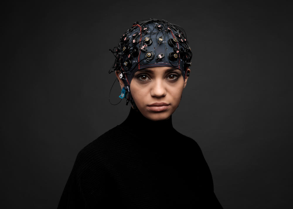
| Works with all types of active and passive biosignal electrodes (g.LADYBbird, g.SCARABEO, g.SAHARA, g.Pangolin, cortiQ electrodes) | |
| Perfectly suited for closed-loop neuromodulation experiments | |
| g.HIamp RESEARCH Edition available (non-certified) |
| Size | 197 (L) × 197 (W) × 90 (H) mm |
| Weight | 1,875 g |
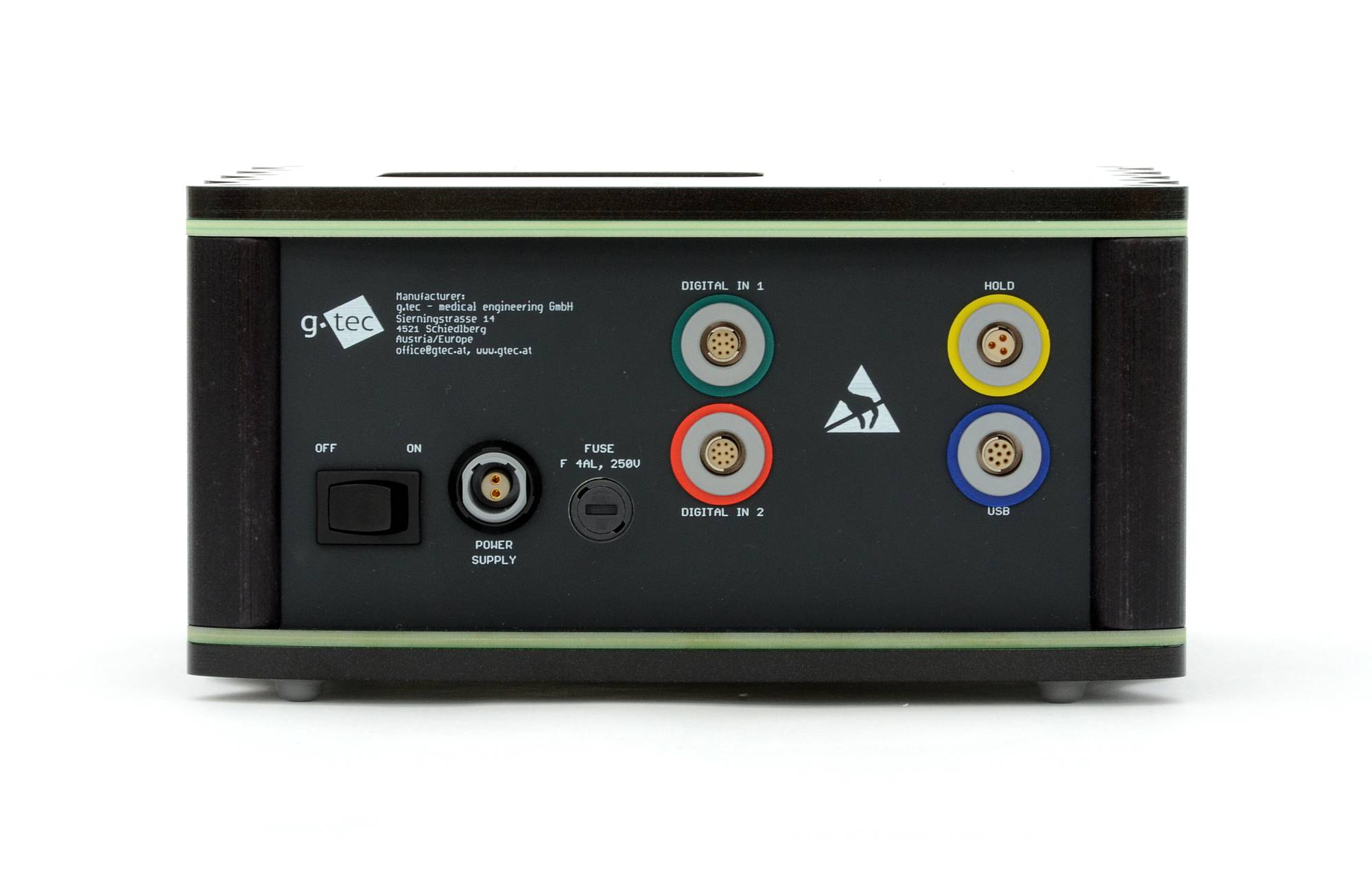
| Interface | USB |
| Digital inputs | 2 x 8 digital trigger inputs 1 x HOLD input (for artifact suppression) |
| Supply | 5V DC, medical mains power supply |
| Sensitivity | 8,57 nV / +/- 340 mV with g.Pangolin |
| Noise level | <0,5 µV rms 1-30 Hz |
| Amplifier type | real DC coupled |
| 256 × ADC | 24 Bit (38.4 kHz internal sampling per channel) |
| DAC | calibration signal |
| Input channels | 256 mono-polar / 128 bi-polar (per device, software selectable) |
| Input impedance | >1000 GOhm // 220 pF |
| Input connectors | standard safety connectors for passive electrodes, 2-pin connectors for active electrodes |
| Applied part | CF |
| Safety class | II |
| Certification and Standards | FDA cleared and CE certified medical product EN60601-1, EN60601-1-2, EN60601-2-26, EN ISO 14971 |
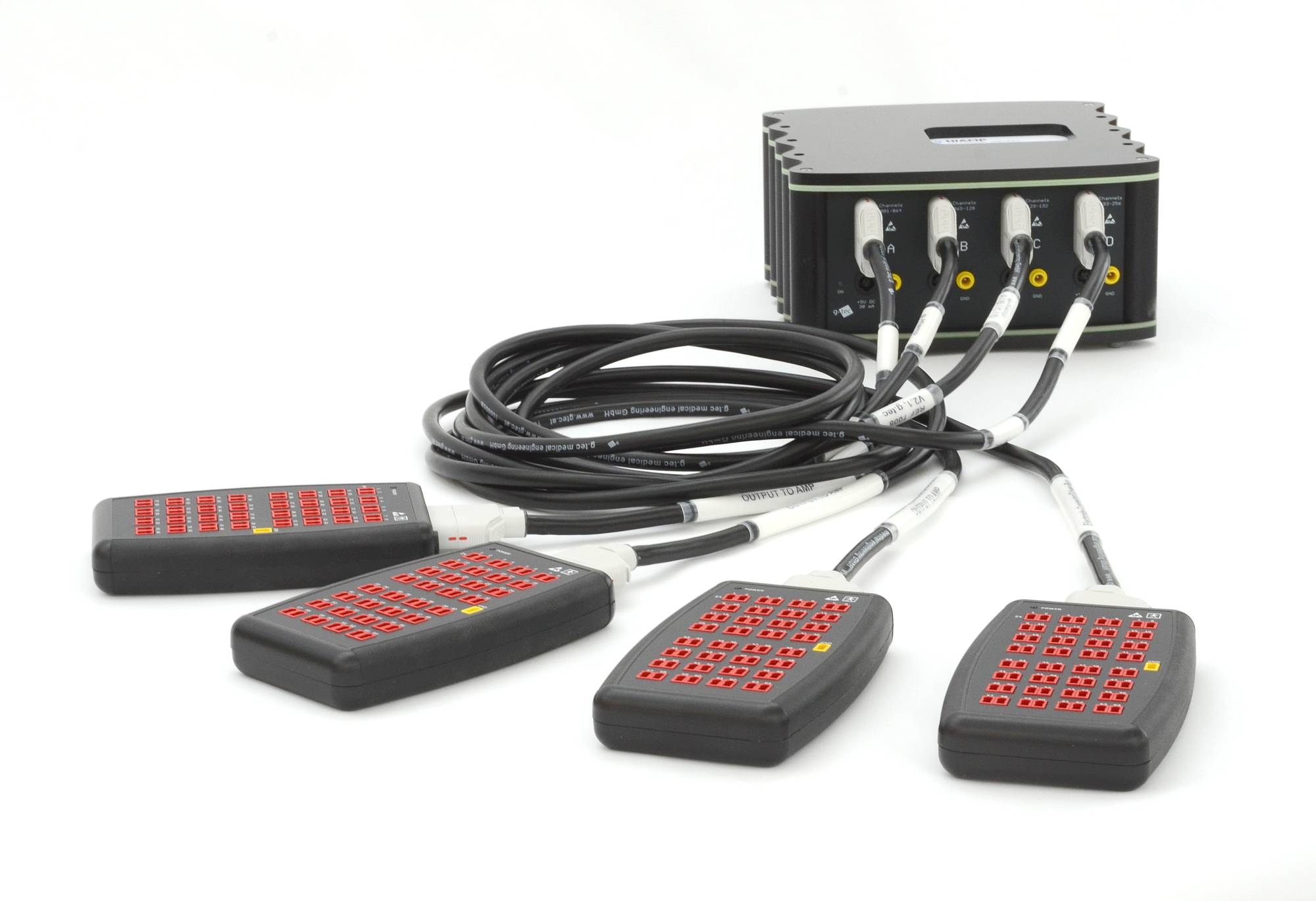
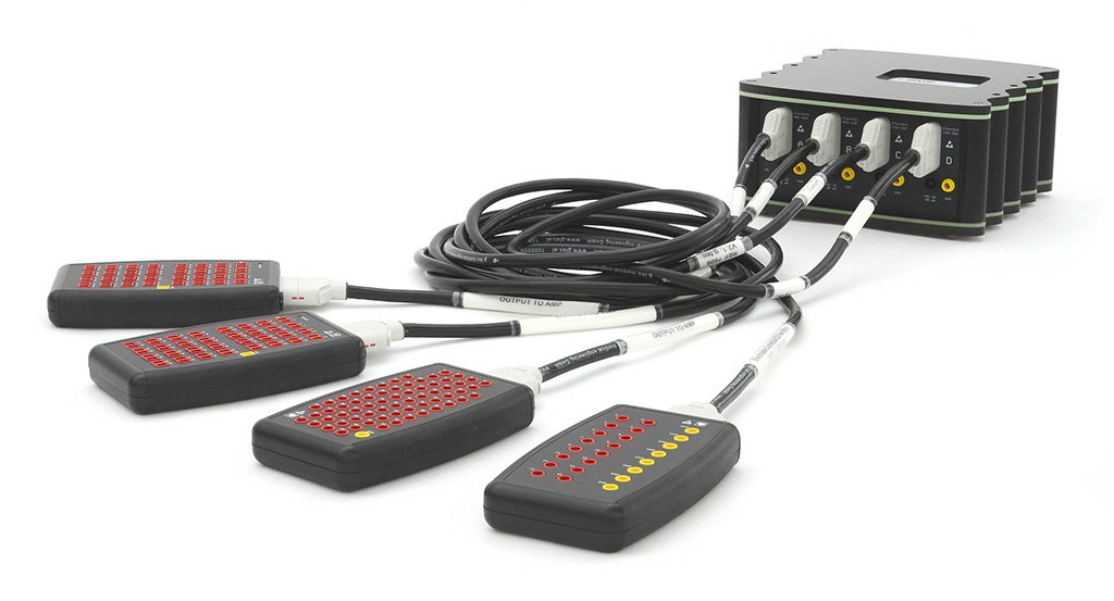
ACTIVE/PASSIVE ELECTRODE CONNECTOR BOXES
A useful feature of the g.HIamp biosignal amplifier is that the main amplifier can be used with all types of g.tec’s EEG electrodes (g.SAHARA, g.SCARABEO, g.LADYbird, g.PANGOLIN, cortiQ electrodes). For that reason, the g.HIamp is highly applicable for all types of neuroscience applications and simultaneous recordings of EEG and TMS/tDCS or fNIRS.
- 64 Channel Passive Electrode Connector Box
- 16 Channel Passive Electrode Connector Box
- 64 Channel Active Electrode Connector Box
- 64 Channel Active Electrode Connector Box TMS
- 64 Channel Passive Electrode Connector Splitter Box
- 256 Channel Active Electrode Connector Box g.Pangolin

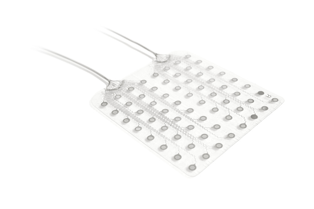
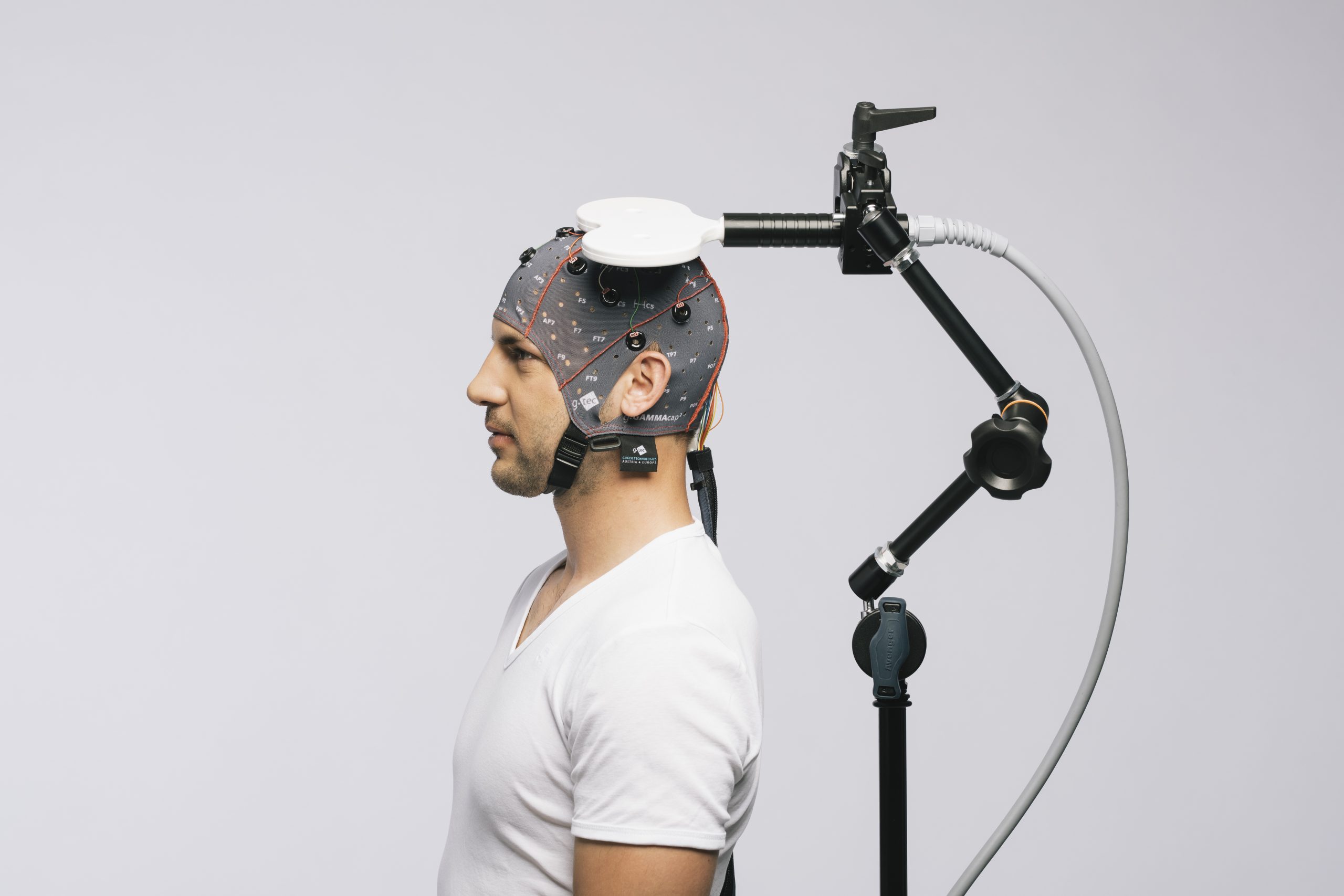
RECORDING ULTRA HIGH-DENSITY EEG
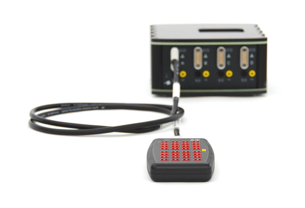
The g.Pangolin ultra high-density EEG electrodes can be used with g.HIamp biosignal amplifier to record high-resolution EEG. The g.Pangolin grids are connected with an active headstage amplifier to the g.HIamp. The g.Pangolin grids have 16 electrodes on a polygon shaped grid that can be mounted onto the head of the person. With the g.Pangolin, up to 1.024 EEG channels can be recorded from the human head to get EEG data with never seen spatial resolution.
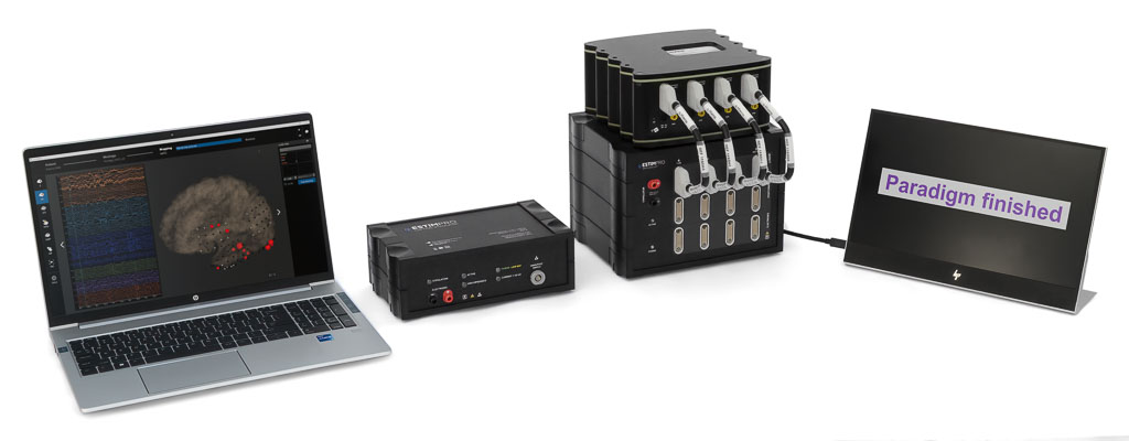
NEUROMODULATION APPLICATIONS
Neuromodulation applications are easily setup with the versatile capabilities of the g.HIamp Multi-Channel Amplifier. This advanced device provides the necessary amplification and signal processing functionalities for precise neural recordings and stimulation. When combined with the g.Estim PRO and it’s Switching Unit, users gain a high-performance neuromodulation setup. The g.Estim PRO facilitates controlled electrical stimulation, allowing for targeted modulation of neural activity. Meanwhile, the g.Estim SWITCHING UNIT adds an essential layer of functionality by enabling seamless sensing, switching, and stimulation of specific brain regions.
RECORDING ECOG
ECoG electrodes are for invasive neuromonitoring and brain-computer interface experiments and it allows to record a high-quality Electrocorticogram (ECoG). It supports brain mapping as well as the monitoring of electrical brain signals, e.g. for the localization of epileptogenic foci, to map the eloquent cortex or to run BCI real-time experiments for a short implantation period less than 30 days.
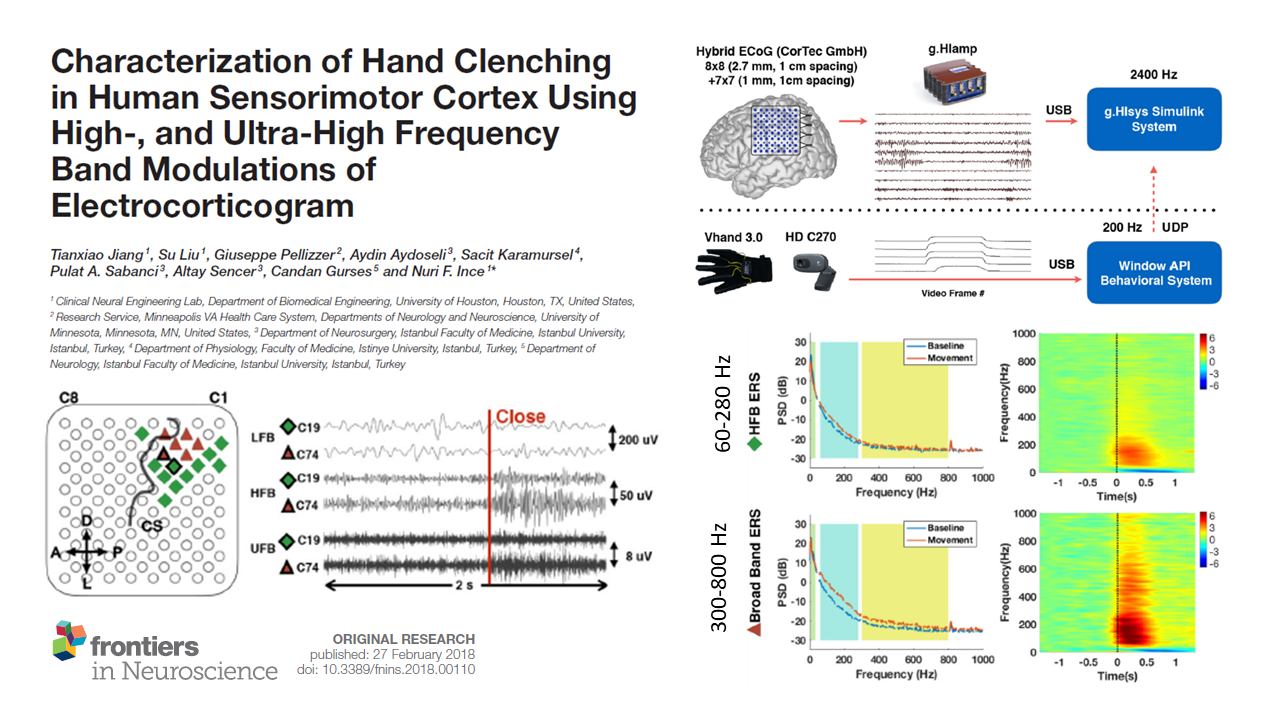
RECORDING HIGH-GAMMA UP TO 1 kHz
Clinical recording systems often can’t capture brain signals above 80 Hz, restricting vital research in neuroscience and brain-computer interface (BCI) studies. Unlike most systems, the g.HIamp breaks this barrier by recording high-gamma brain activity up to 1 kHz. Its specialized design—featuring high oversampling, precise filters, superior amplifiers, and an extensive input range from DC to 6.8 kHz—enables it to detect these crucial signals. It’s essential not to miss these high-frequency components in your experiments. The image illustrates an ECoG modulation experiment conducted by Nuri Firat Ince’s team at the University of Houston, utilizing the g.HIamp during finger stimulation (PDF). Choosing the right equipment can save valuable time for both patients and researchers.

FULL SOFTWARE ENVIRONMENT
g.tec provides a broad spectrum of user-ready and customizable software solutions for different groups of users (e.g. engineers, researchers & scientists, physiologists, medical staff, along with software developers & programmers). g.HIamp is integrated into the main core of BCI2000 and OpenVIBE and supports the LSL (lab streaming layer) as well as a plugin for Dewesoft X that allows you to acquire data with g.HIamp biosignal amplifier inside the Dewesoft data acquisition system for automotive applications.

We are using g.tec’s g.HIamp and we are very happy with it! We are decoding speech which requires perfect synchronization and this is what we achieve with this fantastic device!
Alex Ossadtchi, PhD - Director of Center for Bioelectric Interfaces, Moscow
“Fused with a variety of rapid prototyping and research software tools, g.HIamp serves as a unique tool in our clinical research applications to record electrocorticogram and local field potentials. The oversampling process executed by the internal DSP provides exceptional SNR and enables capturing higher frequency brain rhythms with superior quality.”
Nuri Firat Ince, PhD - University of Houston, USA
“g.HIamp allows us to perform robust intraoperative recordings to ensure cortical and subcortical electrodes are placed in the correct functional regions, which is critical various clinical applications and research directions at the University of Florida Hospital.”
Aysegul Gunduz, PhD - University of Florida, USA
“The g.tec g.HIsys environment allows my lab to rapidly develop new applications. Biomedical engineering students here at Columbia University use the rapid prototyping environment during their education to get familiar with data acquisition, real-time processing and signal analysis.”
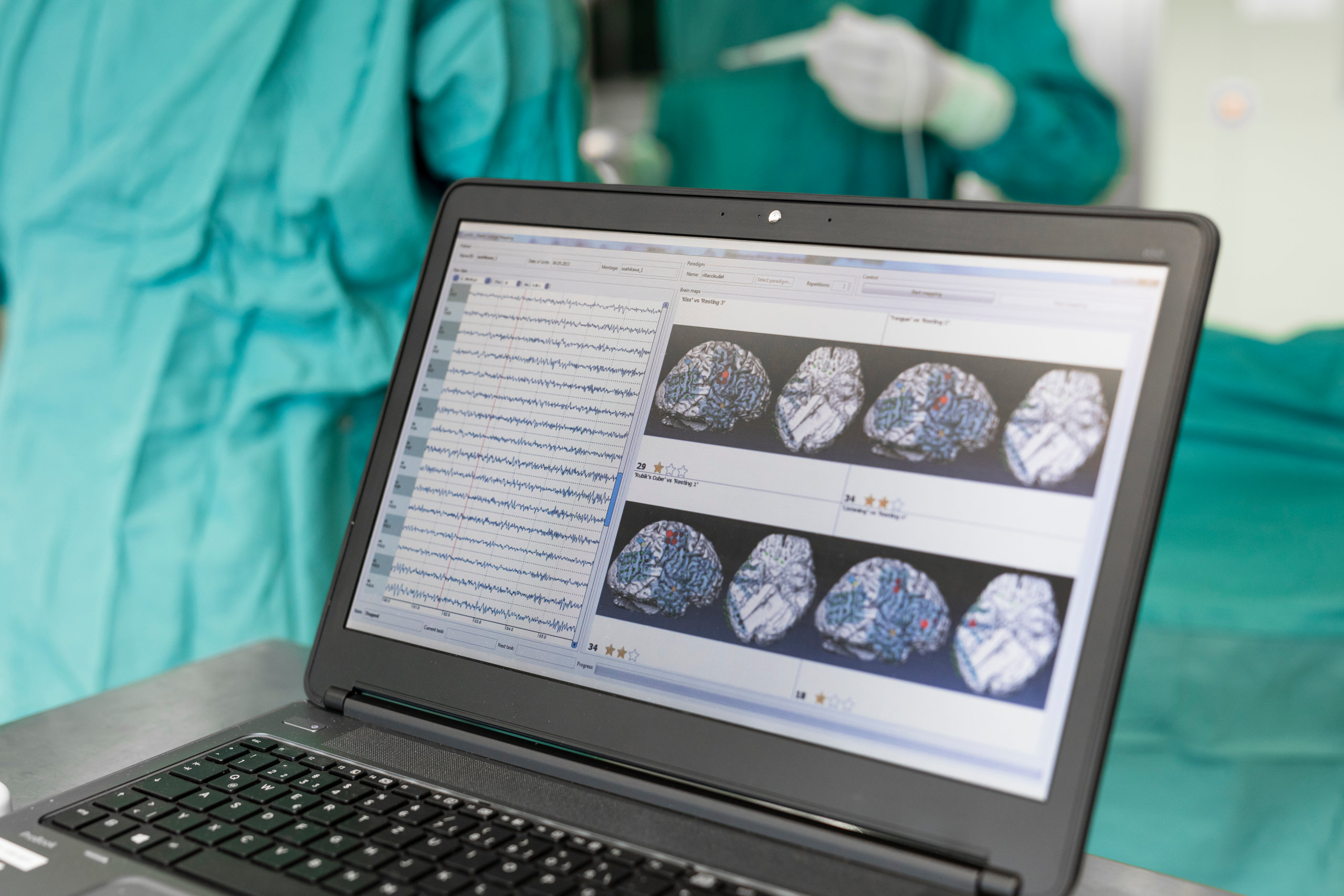
Nima Mesgarani, PhD - Columbia University, New York, USABRAIN MAPPING & EPILEPSY MONITORING
A passive brain mapping procedure based on electrocorticographic (ECoG) signals is a fast and precise mapping technique without the risk of causing pain or seizures. They can accurately identify cortical regions related to receptive and expressive language functions, motor functions and the somatosensory system in the brain.
g.HIamp biosignal amplifier can be used to realize brain mappings in the operating room or neuro monitoring unit.
SIMULTANEOUS TMS & EEG RECORDINGS WITH ARTIFACT REMOVAL ALGORITHM
As a result of the strong electromagnetic field, transcranial magnetic stimulation (TMS) can cause large discharge artifacts in the EEG due to amplifier characteristics and skin/electrode capacitance/impedance. Therefore, it is especially important to use the proper hardware and software. Read this article to learn how to perfectly record TMS and EEG at the same time.
RECORD EEG & FNIRS SIMULTANEOUSLY
g.HIamp in combination with g.SENSOR fNIRS enables simultaneous recordings of EEG and fNIRS (functional near-infrared spectroscopy) signals. A simultaneous recording of both fNIRS and EEG signals is highly beneficial because EEG signals indicate motor imagery while fNIRS signals show long lasting changes like mental counting or pain. In other words, researchers can capture what might be missed when using only EEG or fNIRS.
g.HIamp users can combine g.GAMMAsys technology to record from active g.LADYbird, active g.SCARABEO or g.SAHARA hybrid electrodes with 8 fNIRS sensors by using g.SENSOR fNIRS.
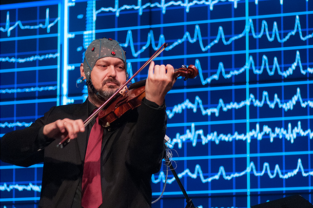
STUDY MUSIC AND THE MIND
The LIVELAB at McMaster Institute for Music and the Mind (MIMM) is engaged in neuroscientific research that aims to understand the positive role of music training, movement and performance. Researchers study music in a live setting using g.HIamp and g.HIsys to learn how performers interact, how audiences move during a performance and the social and emotional impact of these experiences.
THE ULTIMATE RESEARCH EDITION
g.HIamp RESEARCH is the non-certified version of g.HIamp and offers 80/144/256 channels. Therefore, it is less expensive, and intended for research applications only. The device has freely accessible and configurable features, although any kind of ECoG experiments are excluded.
RECORD HIGH-FREQUENCY OSCILLATIONS
High-frequency oscillations (HFOs) can be recorded with the Electrocorticogram (ECoG) with implanted electrode grids in epilepsy patients. The HFOs occur in frequencies between 80-500 Hz. Hence, recording HFOs requires a high-performance biosignal amplifier such as g.HIamp with a high sampling frequency and a very good signal-to-noise ratio and resolution.
HFOs can be found in normal brain structures, but also in pathological networks, and mapping these neural networks is important for pre-surgical planning in epilepsy patients. Therefore, brain surgeons need to distinguish physiological from pathological HFOs. Recently, this was realized by the University of Houston with unsupervised machine learning techniques.
This technology can detect the repetitive waveforms of pathological HFOs within a few minutes, what is an important step for a valid clinical biomarker.
ACCURACY & DATA QUALITY
The amplifier drives each ADC at 614,4 kHz, which is much higher than the required sampling frequency. Then, the floating-point DSP internally performs the oversampling and averages samples to increase the signal-to-noise ratio. The floating-point DSP also performs real-time bandpass filtering and notch filtering of the data. Several hundred different bandpass filters are predefined.
Bipolar derivations can be calculated by the DSP to work with a very high CMRR. g.HIamp uses two additional co-processors for a steep anti-aliasing filter that guarantees a superior signal-to-noise ratio.
All these features together make it possible to record even high-gamma activity in the kHz range! The amplifier uses 256 ADC for the 256 channels, and hence all signals are sampled exactly at the same time point to avoid any time delay between channels. This is especially important for brain mapping procedures.
INPUT CHANNEL PROPERTIES
g.HIamp uses wide-range DC-coupled amplifier technology in combination with 24-bit sampling. The result is an input voltage of +/- 340 mV with a resolution of 85,7 nV! This means that every electrophysiological signal can be recorded directly, without additional hardware.
Neither high electrode offset voltage nor large artifacts resulting from electrical or magnetic stimulation will saturate the amplifier inputs. This feature is important for various artifact treatment and correction algorithms.
The use of digital filters avoids hardware related variations between channels.
CHANNEL COUNT
g.HIamp can be ordered with 64+16, 128+16 or 256 channels. The 64+16 channel version comes with one 64 and one 16 channel connector box. The 128+16 version comes with two 64 channels and one 16 channel connector box, and the 256 channel version comes with four 64 channel boxes.
If the multi-device toolbox from g.HIsys is used up to 1024 channels can be recorded.
SKIN-ELECTRODE IMPEDANCE
g.HIamp uses a new principle for impedance measurement that can determine the skin-electrode impedance for both passive and active electrodes. It works for both gel or dry electrodes provided by g.tec, and even for ECoG grids.
The impedance values are color coded, and all 256 channels are shown in one window, which is updated every few seconds! The impedance check can also beep if an electrode was successfully mounted. This allows very fast assembly of electrodes during real-time impedance control.
NEED MORE INFORMATION
ABOUT THIS PRODUCT?
Send us your email so we can contact you as soon as possible.


