


PROGRAMMABLE ELECTRICAL STIMULATION FOR FUNCTIONAL BRAIN MAPPING & CLOSED-LOOP RESEARCH
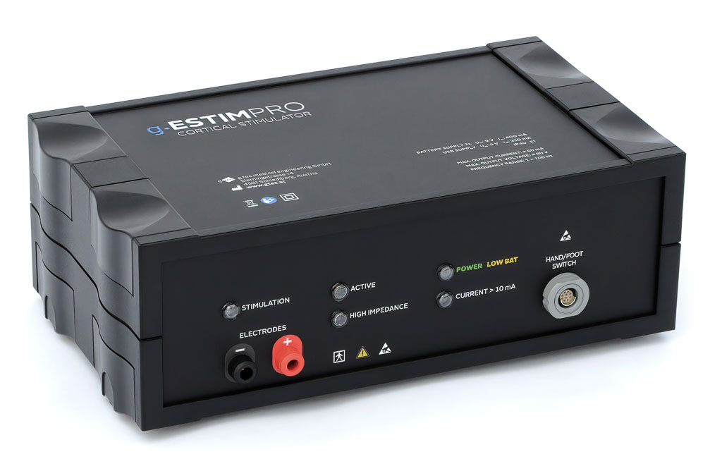
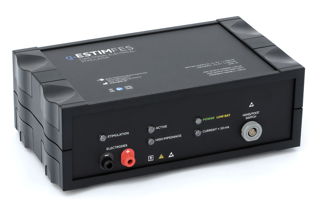
g.Estim PRO is a programmable constant-current cortical stimulator designed for electrical stimulation of neural tissue during functional brain mapping procedures. It delivers configurable biphasic stimulation pulses and is intended for applications such as electrical cortical stimulation (ECS) mapping prior to cortical resections near functionally essential brain areas.
The system is CE-certified in Europe and FDA-cleared in the United States for use in human patients and must be operated by medically trained and qualified personnel in a medical environment.
g.Estim PRO provides an integrated electrode impedance check and monitors the applied stimulation current and voltage for verification. With a maximum electrode output of ±80 V DC, g.Estim PRO provides the voltage range required to maintain the programmed constant current across varying electrode-tissue impedances within the specified operating limits.
For research applications, g.Estim PRO can be integrated with g.HIamp or g.USBamp and g.HIsys to acquire EEG/ECoG signals, analyze brain activity in real time, and control cortical stimulation within closed-loop experiments. The research software upgrade provides programmatic access for adapting stimulation parameters during the experiment.
| Programmable cortical stimulation | Biphasic constant-current electrical stimulation |
| 0.2–15 mA stimulation current | Configurable in 10 µA increments |
| Maximum electrode output ±80 V DC | High output-voltage capability for constant-current stimulation |
| Flexible stimulation parameters | Configure current, phase duration, pulse rate and stimulation trains |
| Impedance & stimulation monitoring | Integrated electrode impedance check and monitoring of applied stimulation |
| Digital synchronization | Trigger outputs for synchronization with other experimental devices |
| Real-time research control | API-based control for programmable and closed-loop stimulation workflows |
| Clinical regulatory status | CE-certified and FDA-cleared for specified clinical use |
| Stimulation type | Biphasic constant-current stimulation |
| Stimulation current | ±0.2–15 mA |
| Current resolution | 10 µA |
| Current accuracy | ±10% or ±50 µA, whichever is greater |
| Phase shape | Rectangular |
| Phase duration | 0.1–1.0 ms |
| Phase duration resolution | 10 µs |
| Pulse rate | 2–100 pulses/s |
| Pulse rate resolution | 0.1 pulses/s |
| Pulse onset interval | 10–500 ms |
| Train duration | 1 pulse–20 s |
| Maximum electrode output | ±80 V DC |
| Power supply | 2 × 9 V batteries; USB connection |
| Standards | IEC 60601-1, IEC 60601-1-2, IEC 60601-2-40, IEC 62304, IEC 62366, ISO 14971 |

STIMULATE, SWITCH & RECORD
Extend g.Estim PRO into an integrated cortical stimulation and electrophysiological recording system with the g.Estim PRO Switching Unit and g.HIamp.
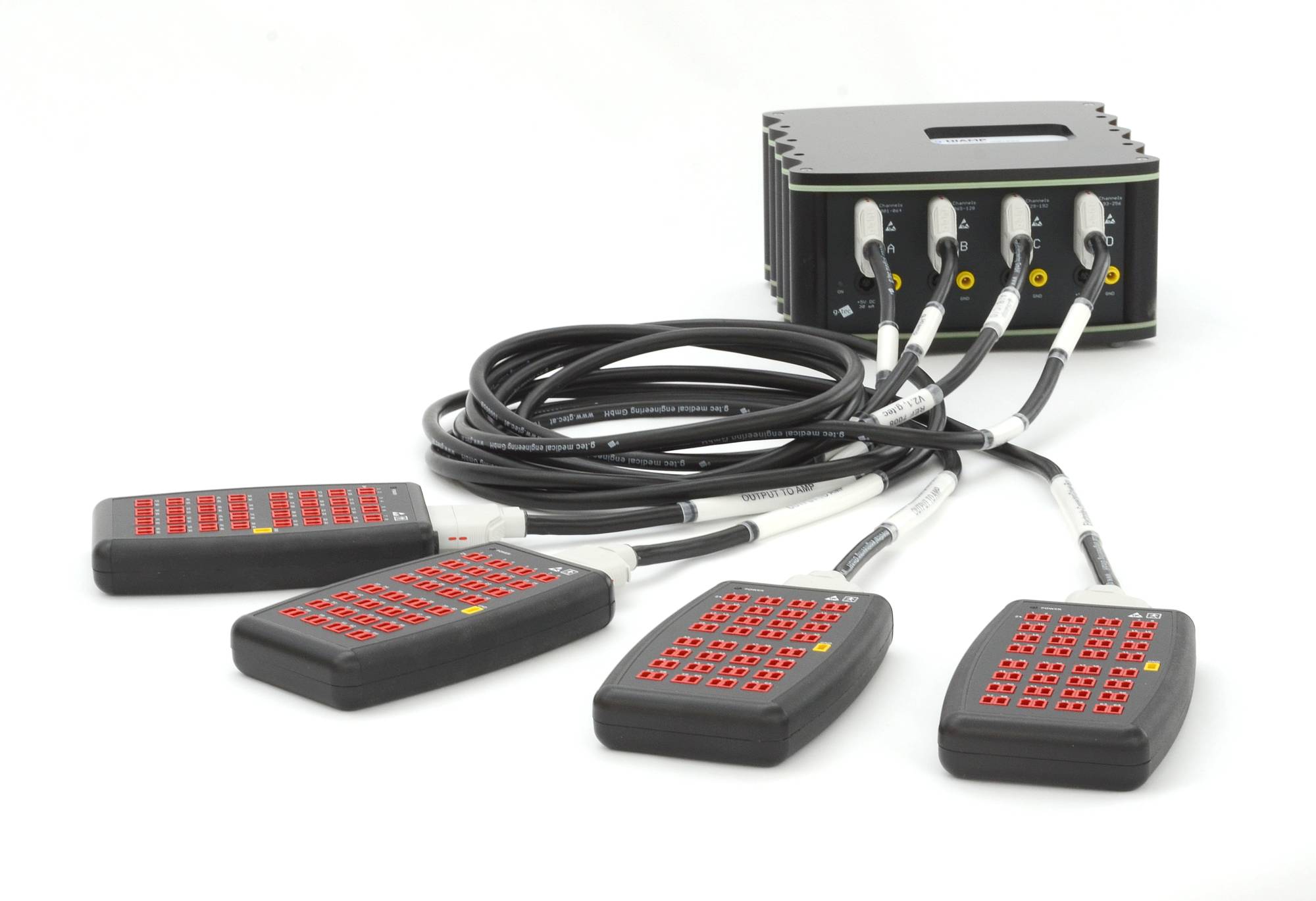
The Switching Unit provides programmable electrode routing for up to 256 channels, allowing selected cortical electrodes to be routed between stimulation and recording pathways. Combined with g.HIamp, the system enables cortical activity to be recorded between and during stimulation for functional brain mapping and advanced stimulation research.
The Switching Unit also supports advanced impedance analysis from 20–1000 Hz across a measurement range of 1–800 kΩ.
REAL-TIME & CLOSED-LOOP CORTICAL STIMULATION
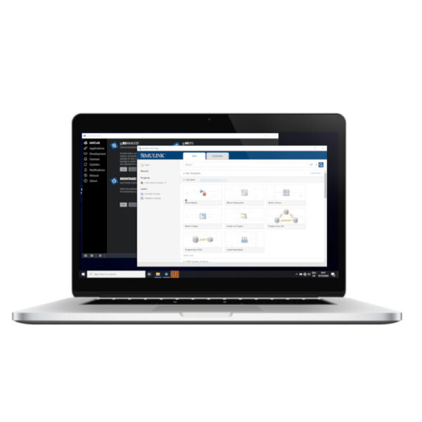
For advanced research applications, g.Estim PRO can be integrated with g.HIamp or g.USBamp and g.HIsys to create real-time and closed-loop stimulation workflows. EEG or ECoG signals can be acquired and analyzed in real time, and extracted signal parameters or classification results can be used to control cortical stimulation. The g.Estim PRO research software upgrade provides programmatic control of stimulation parameters for experiments that dynamically link recorded brain activity with electrical stimulation. g.HIsys supports direct control of g.Estim PRO from the Simulink environment.

FLEXIBLE STIMULATION CONTROL
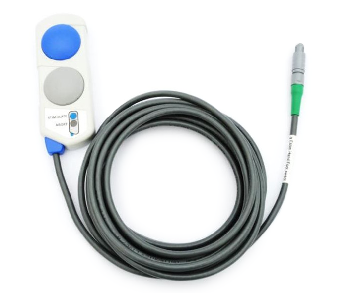
g.Estim PRO can be controlled from the computer or combined with dedicated hand and foot switches for direct stimulation control during functional brain mapping procedures. The Hand Switch provides manual control close to the stimulation site, while the Foot Switch enables hands-free operation, allowing the operator to choose the control method that best fits the clinical workflow.
ADVANCED IMPEDANCE MEASUREMENT
The g.Estim PRO Switching Unit delivers high-precision impedance measurements, allowing you to assess electrode impedance over a broad frequency (20-1000 Hz) and magnitude range (1-800 kOhm). With the intuitive Impedance Measurement Tool, you can easily configure settings, review results, and generate publication-ready reports.


STIMULATION LOG VIEWER
StimLogViewer provides a comprehensive view of the most important events and parameters from your brain stimulation sessions. Effortlessly review stimulation sites, current amplitudes and timings, system states, and much more – either in real-time during your experiment or by revisiting past sessions.
SIMULINK INTERFACE
Integrate g.Estim PRO directly into MATLAB/Simulink with g.HIsys to build custom stimulation and neuromodulation experiments. Researchers can control stimulation from their Simulink models and dynamically modify parameters such as stimulation amplitude, phase duration, pulse rate, number of pulses, polarity, trigger timing, and stimulation start or abort.
This makes it possible to connect custom signal-processing algorithms, experimental logic and stimulation control within the same real-time research environment, from event-triggered stimulation to advanced closed-loop neuroscience applications.
SEEING FACES AND RAINBOWS THROUGH CORTICAL STIMULATION
What happens when specific regions of the human brain are electrically stimulated? In this experiment, researchers combined ECoG recording with direct electrical cortical stimulation to investigate face- and color-selective regions of the ventral temporal cortex. Stimulation of the fusiform face area produced illusory face percepts known as “facephenes,” while stimulation of nearby color-preferring sites produced rainbow and color percepts. The study provides striking causal evidence for the functional specialization of the human brain.
Watch the video to see how ECoG and electrical cortical stimulation were used to explore the neural mechanisms of face and color perception.

MAP FUNCTIONAL BRAIN NETWORKS WITH CORTICAL STIMULATION
How can we identify not only where a brain function is located, but how functional regions are connected? This lecture demonstrates how electrical cortical stimulation, ECoG, cortico-cortical evoked potentials (CCEPs) and white matter tractography can be combined to investigate functional and anatomical brain networks.
See how g.Estim PRO, g.HIamp and g.HIsys support workflows ranging from functional brain mapping and stimulation-evoked response recording to real-time CCEP analysis and network visualization. The presentation also demonstrates how programmable electrode switching can automate sequential stimulation and recording for systematic cortical network screening.

MEDIAN NERVE STIMULATION & SEP MAPPING
Identify the central sulcus using somatosensory evoked potentials.
Median nerve stimulation produces a peripheral response and a somatosensory evoked potential (SEP) that can be recorded from the cortex. Detecting the characteristic phase reversal of the SEP across the central sulcus provides an important functional landmark for neurosurgical brain mapping.


PERIPHERAL & INTRACRANIAL BRAIN MAPPING
Stimulate the periphery. Stimulate the cortex. Map brain function.
Peripheral nerve stimulation can evoke measurable cortical responses, while direct cortical stimulation can produce or disrupt functional responses. Combining stimulation with ECoG recording enables researchers to investigate sensory and motor function, cortical organization and stimulation-response relationships within integrated brain-mapping workflows.
CLOSED-LOOP DEEP BRAIN STIMULATION RESEARCH
Explore adaptive stimulation based on real-time neural activity.
g.tec biosignal acquisition, real-time processing and electrical stimulation technologies can be combined to investigate closed-loop DBS research. Local field potentials and other electrophysiological signals can be analyzed in real time and used to adapt stimulation parameters, supporting research into stimulation timing, electrode positioning and responsive neuromodulation.


REAL-TIME ECOG & DIRECT CORTICAL STIMULATION FOR LANGUAGE MAPPING
Noll et al. – Neuro-Oncology Practice, 2024
Researchers at MD Anderson Cancer Center demonstrated an intraoperative language-mapping workflow combining real-time ECoG with direct cortical stimulation (DCS) during awake brain surgery. The system used g.Estim PRO for biphasic cortical stimulation, the g.Estim PRO Switching Unit for switching between recording and stimulation, and the 256-channel g.HIamp for high-resolution ECoG acquisition. g.HIsys and MATLAB/Simulink provided real-time signal acquisition, processing and visualization.
Real-time gamma-band ECoG mapping identified a language-related cortical region that corresponded with the direct cortical stimulation results: stimulation of the same electrode region at 6 mA produced consistent speech arrest during object and action naming tasks. The authors concluded that real-time ECoG visualization is feasible and may help identify targets for direct cortical stimulation during intraoperative language mapping.
REVEALING PATHOLOGICAL BRAIN NETWORKS WITH CCEP
Kamada et al. – Frontiers in Neurology, 2020
Kamada et al. investigated functional and pathological brain networks in 14 patients with intractable epilepsy using electrical stimulation, cortico-cortical evoked potentials and high-gamma mapping. Intracranial recordings were performed with a 256-channel g.HIamp, while electrical stimulation at different cortical sites was used to identify functionally connected brain regions.
The study showed how stimulation and high-channel-count electrophysiology can reveal pathological network connectivity and support surgical decision-making. 13 of 14 patients (92.8%) who underwent network surgery had a favorable prognosis, demonstrating the clinical relevance of combining stimulation, CCEP recording and functional mapping.


MAPPING LANGUAGE NETWORKS WITH ELECTRICAL STIMULATION
Tamura et al. – Journal of Neurosurgery, 2016
Tamura et al. combined real-time high-gamma mapping with electrical cortical stimulation and cortico-cortical evoked potentials (CCEP) in five patients undergoing awake brain tumor surgery. Electrical stimulation of identified receptive language regions evoked measurable responses in connected frontal language areas, demonstrating how cortical stimulation and intracranial recording can be combined to investigate functional language networks.
For electrocortical stimulation mapping, the researchers applied biphasic pulses at 50 Hz with a 0.3 ms pulse duration, starting at 3 mA and increasing the stimulation intensity when required up to a maximum of 15 mA.
FREQUENTLY ASKED QUESTIONS
g.Estim PRO includes an integrated electrode impedance check and monitors the applied stimulation current and voltage, allowing stimulation conditions to be verified during use. Stimulation sessions can additionally be reviewed with StimLogViewer, including stimulation sites, current amplitudes, timing and system states.
g.Estim PRO provides digital outputs for synchronization with external equipment. In integrated g.tec workflows, stimulation can be synchronized with electrophysiological acquisition using g.HIamp or g.USBamp. This enables stimulation events to be related to recorded EEG/ECoG activity and incorporated into real-time experimental workflows.
Yes. g.Estim PRO can be combined with the g.Estim PRO Switching Unit and g.HIamp to create an integrated stimulation and electrophysiological recording system. The Switching Unit provides programmable electrode routing for up to 256 channels, allowing selected cortical electrodes to be routed between stimulation and recording pathways. The setup enables cortical activity to be recorded between and during stimulation.
This architecture has also been used in published intraoperative research. Noll et al. used g.Estim PRO, the Switching Unit, a 256-channel g.HIamp and g.HIsys in a real-time ECoG and direct cortical stimulation language-mapping workflow.
The Switching Unit completes electrode switching within 6 ms after a digital trigger and provides a digital output indicating the switching state, enabling the switching event to be recorded together with the electrophysiological signals.
Yes. The g.Estim PRO research software upgrade provides programmatic control of stimulation parameters. Together with g.HIsys, g.Estim PRO can be controlled from the MATLAB/Simulink environment and integrated with g.HIamp or g.USBamp for real-time EEG/ECoG acquisition, signal processing and stimulation control. Signal parameters or classification results can therefore be incorporated into closed-loop research workflows.
Yes. g.Estim PRO can be integrated with g.HIamp, g.HIsys and the g.Estim PRO Switching Unit for stimulation and intracranial electrophysiological recording workflows used in CCEP and functional connectivity research. Programmable electrode routing allows stimulation and recording configurations to be changed systematically across implanted electrodes, while electrophysiological responses can be acquired and processed within the same g.tec ecosystem.
g.Estim PRO is a programmable constant-current cortical stimulator with a stimulation range of 0.2–15 mA, 10 µA current resolution and an accuracy of ±10% or ±50 µA, whichever is greater. It provides a maximum electrode output of ±80 V DC and rectangular stimulation phases from 0.1–1.0 ms in 10 µs increments, with an accuracy of ±10% or ±20 µs, whichever is greater. Pulse rates can be configured from 2–100 pulses/s with 0.1 pulses/s resolution, pulse onset intervals from 10–500 ms, and train durations from 1 pulse to 20 seconds. The inter-phase duration is 0 ms.
The voltage required to maintain the programmed current depends on electrode-tissue impedance according to U = I × R. g.tec recommends an electrode-tissue impedance below 5 kΩ. For loads below 5 kΩ, specified rise and fall times are <5 µs; for loads between 5 and 800 kΩ, the specified relationship is tr = tf < Rload × 1 ns.
g.Estim PRO provides two digital outputs, DO1 and DO2, for synchronization with g.HIamp, g.USBamp and compatible external equipment. In g.HIsys research workflows, digital outputs can be configured for TRAIN, PULSE or PHASE triggers, with programmable pre- and post-trigger timing. Hardware-triggered stimulation of up to 250 Hz is supported in the documented g.HIsys workflow.
With the g.Estim PRO Switching Unit, a rising or falling digital-input edge results in completed electrode switching within 6 ms, while the minimum Active State is 1 ms. In the documented g.HIsys Fast Stimulation configuration, the shortest achievable combined switching and stimulation-onset delay is approximately 13 ms. For Stimulate-and-Record workflows, a pre-trigger of at least 6 ms allows electrode switching to complete before stimulation begins.
The Switching Unit supports up to 256 electrode channels, a maximum stimulation voltage of 80 V, maximum stimulation current of 15 mA and maximum stimulation frequency of 500 Hz.
g.Estim PRO provides DO1 and DO2 digital outputs with high-level voltages of 2.4–3.3 V DC and low-level voltages of 0.0–0.4 V DC. DI1 uses high-level voltages of 2.0–5.0 V DC and low-level voltages of 0.0–0.8 V DC. Trigger-output cables use a push-pull connector to BNC and have a length of 1 m. The PC interface is USB with an operating range of 4.75–5.25 V DC.
For research applications, g.HIsys provides MATLAB/Simulink integration and the
gEstimPRO_ParameterUpdate()
function. Programmatically controllable parameters include phase amplitude, phase duration, pulse rate, number of pulses, polarity, modality, fade-in/fade-out parameters, digital-output mode, pre- and post-trigger timing, electrode surface area, Start/Abort and Active/Stop. Most parameter changes are transmitted immediately and become effective with the next stimulation, while phase amplitude and phase duration can take effect immediately under the documented conditions.
For the Switching Unit, the digital input uses 2.0–3.3 V high and 0.0–0.8 V low, with a minimum trigger duration of >200 µs. Its digital output indicates the active switching state and can be recorded by g.HIamp for precise synchronization.
In g.HIsys ECS workflows, separate post-processing files can be generated for each stimulation site containing the acquired electrophysiological data, stimulation-site information, stimulation-onset triggers and stimulator state. StimLogViewer can additionally be used for reviewing stimulation-session information.
g.Estim PRO is a Class II medical electrical device with a Type BF, galvanically isolated applied part, IP40 protection and S1 continuous operating mode. EMC compliance includes IEC 60601-1-2, and the device requires periodic technical inspection every 12 months according to EN 62353:2014.
Operating conditions are +10 to +35 °C, 20–80% relative humidity, non-condensing, and 800–1060 hPa atmospheric pressure. Transport and storage conditions are −20 to +60 °C, 10–95% relative humidity and 700–1060 hPa.
The system uses 2 × 9 V lithium batteries plus USB, with rated power consumption of 1.75 VA via USB and 3.6 VA via battery. The documented PC configuration includes USB 3.0, Windows 11 Pro 64-bit, at least 8 GB RAM, more than 100 GB storage and an Intel processor with at least two cores at 2.30 GHz.
g.Estim PRO is CE marked, identified as a prescription device in the USA, carries UDI information and is classified as Class II electrical protection with a Type BF applied part. The product documentation references IEC 60601-1, IEC 60601-1-2, IEC 60601-2-40, IEC 62304, IEC 62366, ISO 14971 and EN 62353.
For simultaneous cortical stimulation and electrophysiological recording, the g.Estim PRO Switching Unit additionally specifies a ±340 mV channel input range, AC coupling from 0.16–70 kHz, ≤−70 dB channel crosstalk, ≤6 µV peak-to-peak input-referred noise from 0.5–50 Hz at 256 Hz sampling, typical noise of <1 µV RMS from 1–30 Hz, and calibrated channel offset of <±50 µV.
For tenders, I would still add EU MDR classification, CE/Notified Body certificate identifier, FDA 510(k) number, Basic UDI-DI, device dimensions, weight and battery runtime once these values have been confirmed internally.

NEED MORE INFORMATION
ABOUT THIS PRODUCT?
Send us your email so we can contact you as soon as possible.


