
A Practical Guide for Combined fNIRS & EEG Recordings
Combined fNIRS and EEG recordings become more popular because their complementary approach extracts much more information from the brain than one single technique. But in order to conduct a successful measurement, some key factors have to be considered. Here is a practical guide based on more than 20 years of experience, so that you – as a researcher or medical professional – learn what’s important to perfectly record brain activity and to save time, money and nerves 🙂 Let’s get to it!
What’s the difference between fNIRS vs. EEG?
fNIRS (functional near infrared spectroscopy) measures the oxygenation level and hemodynamic of the brain non-invasively. The oxygenation level changes as brain areas become more active. Researchers can identify brain activity in real-time based on changes in blood oxygenation and other factors. fNIRS is measured with optodes, using LEDs or lasers to emit light into the brain and the reflected light is measured with a detector. By looking at a certain wavelength of the light, the absorption of the hemoglobin can be measured and the blood flow in certain brain regions can be identified. These optodes have normally a distance of 20-30 mm from each other and the fNIRS signal measures the blood flow in between these optodes.
In contrast, the EEG (electroencephalogram) measures the electrical activity of the neurons right underneath the electrodes itself and provides high temporal resolution, unlike fNIRS.
However, simultaneous recording of both fNIRS and EEG signals in one single device is highly beneficial. EEG signals can identify short activities like evoked potentials, while fNIRS signals show long lasting changes like mental counting or pain. In other words, researchers can capture what might be missed when using only EEG or fNIRS.
How can I combine fNIRS and EEG in one single recording?
To measure both EEG and fNIRS at the same time, it makes sense to put the EEG electrodes in between the optodes, because EEG electrodes are smaller and easier to place. Then the EEG electrode picks up the electrical activity at the same spot as the fNIRS picks it up.
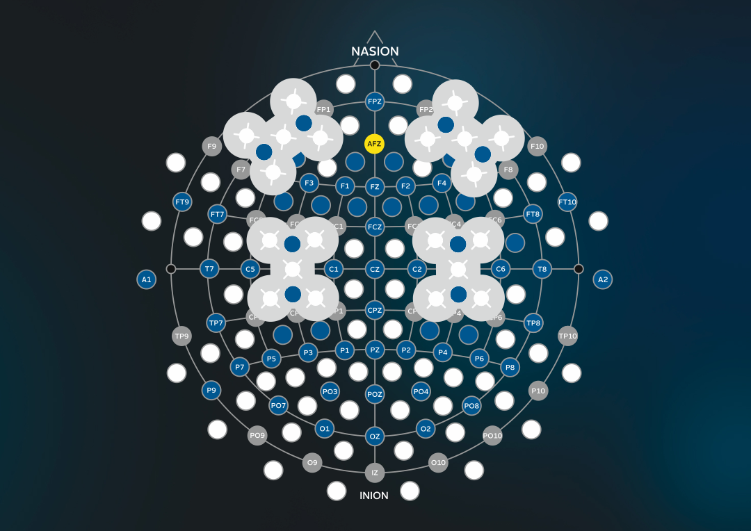
An important factor is the electrode cap. The EEG cap must be able to keep the fNIRS optodes and EEG electrodes in a fixed position to get artifact free data. The dark electrode cap material ensures that no distortions are produced by ambient light. Furthermore, it’s important that both the EEG electrodes and fNIRS optodes are lightweight to reduce movement artifacts in the recordings. Besides that, active EEG electrodes help researchers to get top-quality EEG recordings from 64/32/16/8 g.SCARABEO EEG channels and 8 fNIRS channels within a few minutes. This approach is becoming more and more popular because combining two recording types extracts way more information than using one single method.
The wearable g.Nautilus EEG amplifiers, g.HIamp and g.USBamp can be upgraded with a g.SENSOR fNIRS that enables simultaneous recordings of EEG and 8 fNIRS (functional near-infrared spectroscopy) signals from one device only.
Furthermore, g.tec devices are integrated with NIRSport2 from NIRx. Here, the g.GAMMAcap has 8/16/32/64 electrode holder rings to use g.SCARABEO electrodes and 32 optode holder rings for 16 sources and 16 detectors. This allows you to arrange the detectors and sources in various different ways for having more fNIRS channels.

What equipment is suitable for combined fNIRS EEG recordings?
g.tec has the ideal equipment: You can choose between several EEG and biosignal amplifiers, active wet or hybrid dry EEG electrodes, different sizes of EEG caps and a software solution to quickly set up your neuroscience experiment. Use a g.Nautilus wearable EEG, g.USBamp and g.HIamp biosignal amplifier to measure high-quality EEG signals together with the g.SENSOR fNIRS or with NIRSport2. The EEG electrodes and the fNIRS sensors are assembled in the EEG cap to have well defined positions and proper contact to the scalp.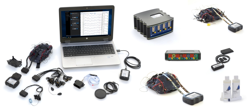
g.tec’s EEG amplifiers can stream the data in real-time into a computer for real-time analysis so they can use BCIs or run other closed-loop experiments. In this case, both the EEG and fNIRS must be sampled simultaneously with precise real-time data management. The EEG amplifier normally has the higher sampling frequency and therefore acts as the “master device”. The g.HIsys Professional software environment has many examples for running motor imagery BCI experiments or performing mental performance tests such as the STROOP word test.
Pick a Biosignal Amplifier
Choose EEG electrodes
Add a g.SENSOR fNIRS to it
-
- g.SENSOR 8 fNIRS Channels
- NIRSport2
Get the right software solution
When are fNIRS+EEG recordings useful?
The combination of fNIRS and EEG is very useful for neuroscience experiments to understand how the brain processes information. This combination can be useful for many applications beyond research, including neuromarketing and monitoring attention, pain, workload, or fatigue. This combination is also useful if you want to boost BCI performance by using both the EEG and fNIRS features as inputs. The EEG has a much higher temporal resolution than the fNIRS. Hence, the EEG is able to record a short lasting event like a single finger movement or recognition of a familiar face, but with the fNIRS it is easy to capture long lasting activity that might be missed with the EEG.
How much time is required to prepare someone with an fNIRS/EEG system?
The preparation time for fNIRS and 32 EEG channels is about 10 minutes with g.tec’s active technology, which is much faster than with passive EEG Electrodes.